Advancements in ophthalmic technology continue to change the way eye care professionals examine and diagnose their patients, and digital imaging solutions are a prime example.
Digital imaging has given doctors the ability to capture images of the human eye with more detail than ever before, allowing them to more effectively identify issues and treat each patient accordingly.
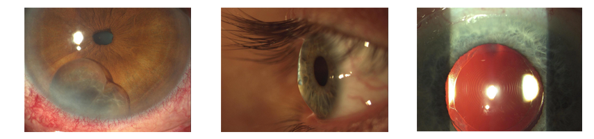
Anterior segment imaging systems in particular are used to capture high-quality contrast images of the front portion of the eye, which includes the cornea, iris, pupil, ciliary body, and lens. This allows for a more efficient diagnosis and better patient education.
How does anterior segment imaging work?
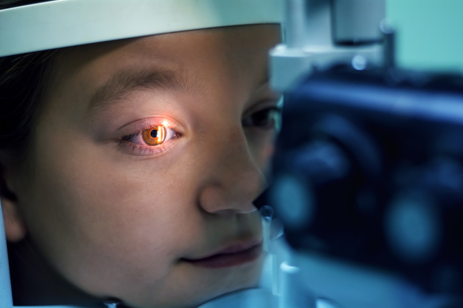
The non-contact imaging method is performed while the patient is in a sitting position. It is convenient and painless, and it requires only a simple wipe-down of the head and chin rests once the exam is complete. This is especially important nowadays as doctors attempt to balance returning to the office with taking necessary safety and distancing precautions to prevent the spread of COVID-19.
Anterior segment imaging systems combine a digital camera with cutting-edge imaging software to capture and store images in an expansive database. With their superior digital resolution, the images can be enlarged, enhanced, segmented, printed, and transmitted to other facilities or ophthalmic professionals over the internet for further analysis.
It also becomes an important diagnostic tool when presented with certain symptoms.
When your patients come to you complaining about symptoms such as blurry vision, dry eyes, contact lens difficulties, headaches, and/or eye pain, digital imaging solutions become an essential part of the exam. Specifically, anterior segment imaging can be crucial for identifying a variety of conditions, including:
- keratitis
- corneal ulcers
- iritis
- uveitis
- corneal abrasion
- cataracts
Because it also pinpoints and quantifies angle closures, anterior segment imaging is a valuable tool for detecting and managing glaucoma, especially angle-closure glaucoma—a leading cause of blindness.
Imaging solutions are also vital for pre- and post-operative exams.
Do you perform refractive surgery or other corneal operations in your practice? Digital imaging plays an important role during the pre- and post-op exam.
Anterior segment imaging can be used to help evaluate the cornea of patients who have had refractive surgery such as LASIK or who have undergone other corneal operations, including phototherapeutic keratectomy (PTK) and corneal transplants.
Why invest in an anterior segment imaging system?
Using the right equipment is vital to the growth and expansion of your practice. Digital imaging technology ensures your patients receive the highest quality of care and sets your practice apart as a leader in ophthalmic wellness.
While all basic eye exams should include use of an ophthalmoscope to look at the outside and back of the eye, as well as a visual acuity exam, not all exams will include the use of an imaging technique. However, when your patients present with more involved symptoms, digital imaging becomes necessary for accurate diagnosis and treatment.
Adding anterior segment imaging to your practice provides several key benefits, including:
- Immediate diagnosis
- Improved patient education
- Expands your ability to serve your patients
- Better collaboration with colleagues
- Increased patient referrals
Digital imaging can also be a new source of revenue for your practice.
Designed to help document pathology, educate patients, and improve collaboration with colleagues, digital imaging can open up a new source of revenue for your practice. In fact, Veatch customers have reported significant increases in revenue by billing for both anterior and posterior images, as a result improving efficiency and increasing patient referrals.
Ready to calculate your return on investment?
Use our free Digital Imaging ROI Calculator. Just enter the number of anterior segment images and retinal images you expect to take in a month, plus the fee for each, along with your estimated monthly lease payment. Click the Calculate button and voilà!
Veatch Ophthalmic Instruments is your first and only source for best-in-class digital imaging equipment to fit your needs and budget. Browse our digital imaging packages or sign up for a free 15-minute efficiency evaluation. If you have any questions or are unsure what system would best suit the needs of your practice, call us at 800.447.7511 to speak to one of our expert account executives.