
In just under two decades since its widespread introduction into the ophthalmic field, optical coherence tomography (OCT) imaging has significantly improved how doctors are able to detect and monitor retinal diseases. Since then, the technology has seen dramatic advancements that have secured its place in the modern eye care practice as a valuable diagnostic tool. In this blog, we will examine briefly the use and benefits of OCT imaging and spotlight an example of a modern OCT camera.
What is optical coherence tomography?
Optical coherence tomography, or OCT, allows eye care professionals to capture high-resolution images of the back of the eye, also called the retina. This non-invasive imaging method utilizes a low-powered, near-infrared laser to photograph the retina and highlight its many layers in order to diagnose and manage a range of conditions that may impact healthy vision.
You will often hear OCT imaging compared with ultrasound technology, the difference being that OCT uses light waves instead of sound waves to photograph the retina and measure its thickness, as well as monitor swelling and fluid. The images also highlight changes in the optic nerve fibers, such as those related to glaucoma, which in turn helps doctors determine whether or not treatment is needed.
Among the conditions and issues that OCT imaging may help detect are age-related macular degeneration (AMD), diabetes-related retinopathy, glaucoma, retinal tear, retinal detachment, macular hole, macular pucker, and macular edema.
What are the benefits of OCT imaging?
The primary benefits of OCT imaging are its powerful 3D resolution and rapid scanning speed.
The higher-resolution images are captured in real time and offer an intricately detailed view of the retina for better overall test results. And, when compared with other diagnostic procedures, OCT imaging equipment delivers faster and more accurate results.
On the patient side, the fact that OCT imaging is non-invasive is a significant benefit. In addition, the cutting-edge technology allows for earlier detection and diagnosis of eye disease, which can help prolong healthy vision.
Beyond ophthalmic and optometric care, OCT imaging technology has myriad applications in healthcare. For example, it is used by cardiologists prior to catheterization to provide images of the blood vessels in and around the heart. It is also used in pulmonology, oncology, dermatology, gastroenterology, and dentistry.
OCT imaging: Spotlight on the Topcon Maestro2
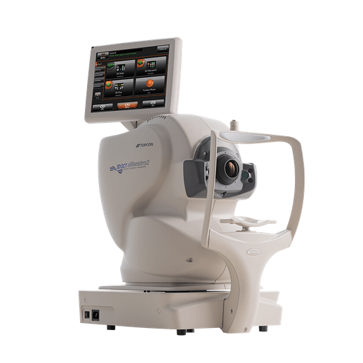
Now available at Veatch Ophthalmic Instruments, the Topcon Maestro2 is a user-friendly OCT fundus camera that automatically performs alignment, focus, and capture with a single touch.
Using the Maestro2, doctors can quickly and effortlessly analyze the retina, optic nerve, and anterior segment with easy-to-interpret reports that can be exported, printed, or sent directly to your EMR in common file formats.
The high-resolution OCT images provided by the Maestro2 offer valuable insight into your patients' eye health by allowing you to detect small changes in the eyes over time and better determine an effective course of treatment. If you would like more information about the Topcon Maestro2 OCT Fundus Camera or other available Topcon products and equipment, contact our team today.