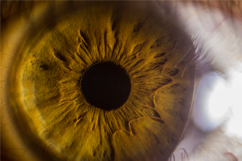
As an eye care specialist, you want to offer your patients the very best in ophthalmic care, and that means outfitting your practice with the latest digital imaging equipment. However, like any new piece of equipment, a digital imaging solution is a significant investment, and you want to make sure you are getting the best return on that investment. Specifically, how can you be sure Medicare will reimburse you for the images you bill for?
While it is true that Medicare reimbursement for anterior segment imaging is not an exact science, there are some things you can do to increase your chances of getting reimbursed. It all comes down to the reason you want to use the technology, and according to Medicare there are only three acceptable reasons for doing so:
- To get additional information that cannot be obtained in any other way during an exam;
- To assess a patient’s disease progress; and/or
- To aid in the diagnosis and treatment of a condition.
We will take a look at each reason in more detail below, but it is important to know that if you perform anterior segment imaging on a patient for any reason other than those listed above, Medicare will likely deny your claim and refuse reimbursement. With that in mind, let’s take a look at the ways in which you can offer anterior segment imaging to your patients and still receive Medicare reimbursement every time you make a claim.
Reason 1: To obtain additional information
If you can prove that you are unable to obtain vital information about a patient’s eye health or vision without using anterior segment imaging, your chances of Medicare reimbursement are good.
Keratoconus is a good example of a condition for which digital imaging is beneficial. Most anterior segment imaging cameras can take images from multiple angles, making the condition easier to spot early on (as it is typically symptom free) and aiding in the diagnosis of keratoconus. Anterior segment imaging as a screening for keratoconus is a must for anyone facing refractive surgery.
And speaking of refractive surgery, smart doctors understand that you cannot always trust the pre-operative test results. Instead, it is a good idea to use anterior segment imaging before you perform or refer any patient for refractive surgery, as well as for monitoring post-operative progress and recovery.
Reason 2: To assess disease progress
Using anterior segment imaging to monitor a patient’s disease progress is one of the most effective ways to receive Medicare reimbursement for the service because a patient with a documented condition almost always automatically qualifies for Medicare reimbursement.
With age-related eye conditions such as dry eye becoming more common among the baby boomer population in particular, anterior segment imaging allows you to more effectively monitor their disease progress and take appropriate steps to help safeguard their vision.
Reason 3: To aid in diagnosis
If you can convince Medicare that you are unable to rule out a serious condition or quantitatively affirm a diagnosis without anterior segment imaging, you will likely be reimbursed for the service. If you truly feel a patient would benefit from anterior segment imaging but have no real grounds for performing the service, citing a suspected condition or potential serious diagnosis will often work in your favor.
However, images alone are not enough to justify your claim.
To increase your chances of reimbursement, you will also need to provide the following to ensure Medicare has enough information to determine whether or not your claim is justified.
- Date of the test
- Order for the images with stated medical rationale
- Reliability of the testing (i.e., patient cooperation)
- Findings from the test
- Diagnosis, if any, based on findings
- Impact of diagnosis on treatment/prognosis
- Signature of the ophthalmologist/physician who performed the test
While there is no guarantee you will be reimbursed, these tips should help.
Digital imaging is highly beneficial for documenting pathology, educating patients, and making your practice more profitable. In fact, our customers have reported significant increases in revenue by billing for both anterior and posterior images, along with better efficiency and more patient referrals.
However, there is never a guarantee that you will be reimbursed, regardless of your documentation and the rationale behind using the service. The best you can do is use these tips to your advantage and document to the best of your ability to ensure your patients receive optimal care and your practice is reimbursed accordingly.