
Back in the day: A History of Imaging Solutions
While there are new developments regularly coming out to ophthalmic photography and imaging solutions, the field has a long history, dating back to the 1800s. Since Dr. Edward Jackson, who wished for tools to record fundus changes over time back in 1886, and Dr. Howe, who took the first photographs of a living human retina in the same year, the field has grown extensively.
For many years, retinal photography was not included as part of a regular exam because of the many challenges and limitations associated with the available methods in that time. Exposure times, daguerreotypes and wet plates, lighting, and processing chemicals at that time made the earliest photographs poor representations of the eye and its interior components and structure.
The same was true around the world. In Portugal, the Netherlands, and other countries, ophthalmologists were beginning to take photos of the eye, but also faced slow film sensitivity, small apertures, and long recharging times.
However, leaders began building on previous tools. These early inventors were both artistically and scientifically minded, able to envision the type of photograph and image that would be necessary for a clear diagnosis, while working through the structure and analysis to lead to that specific image.
The 1960s is when ophthalmic photography really began to grow. With the release of various types of film, fundus cameras, and fluorescein angiography, the field has only grown since then, and continues to develop to produce high-level, in-depth images that can help professionals detect and diagnose various eye conditions and diseases.
Imaging Solutions and Pain Points: How it Works
With so many options for capturing digital images of the eye, ophthalmic professionals are able to observe and scan highly-specific areas of the eye. This includes areas where patients may be experiencing and feeling pain.
It’s important to assess chronic eye pain by learning about its duration, frequency, triggers and quality of pain, as well as determining whether or not it is anterior, posterior, intraocular, or orbital.
While many professionals will make referrals with neurologists, pain specialists, psychiatrists, or anesthesiologists, it’s important to first rule out inflammation, including pseudotumors, uveitis, or iritis, or to make sure there are no foreign particles in the eye. Using advanced imaging techniques can help to rule out any causes of eye pain that may accompany other symptoms, like headaches of all types, including migraines, cluster headaches, hemicranias, or trigeminal neuralgia.
The Top 4 Imaging Solutions
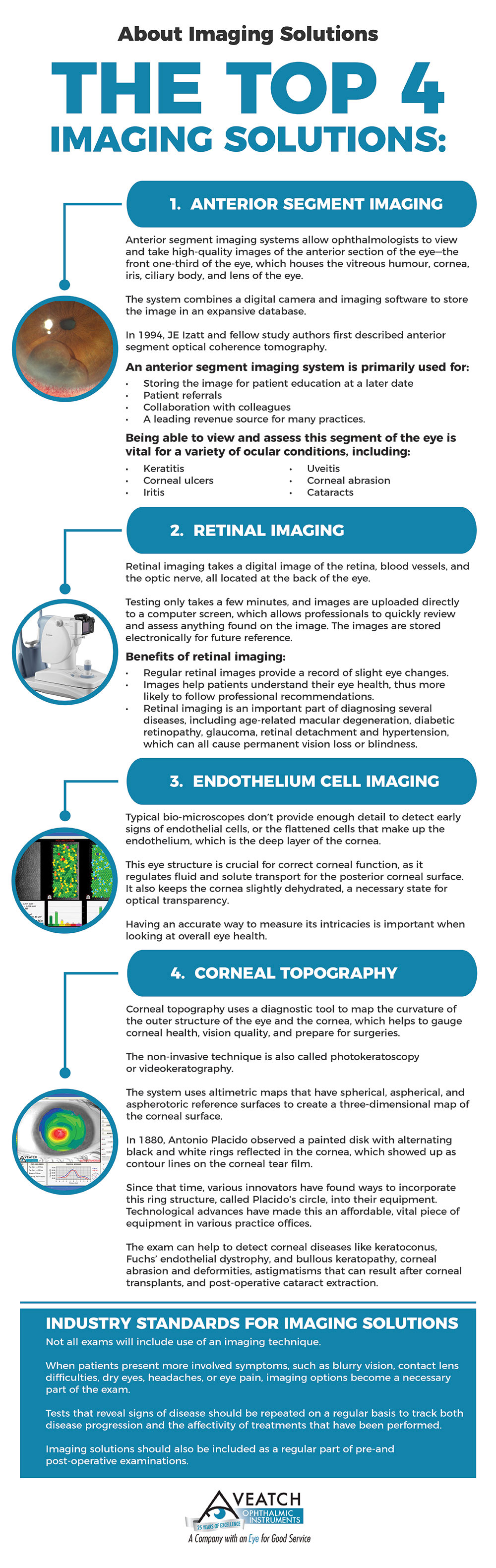
While there are a variety of options that ophthalmic professionals use to diagnose and observe eye diseases, there are four primary solutions that measure different segments of the eye to paint a broad picture of a patient’s overall eye health.
Anterior segment imaging systems allow ophthalmologists to view and take high-quality images of the anterior section of the eye. The anterior segment of the eye is the front one-third of the eye, which houses the vitreous humour, cornea, iris, ciliary body, and lens of the eye. The system combines both a digital camera and imaging software to store the image in an expansive database.
In 1994, JE Izatt and fellow study authors first described anterior segment optical coherence tomography; the field has grown rapidly since that time, with more in-depth wavelengths, real-time imaging, and an increased number of frames per second.
The system has many uses in a practice; primarily, an anterior segment imaging system is used for:
- Storing the image for patient education at a later date
- Patient referrals
- Collaboration with colleagues
- A leading revenue source for many practices.
Being able to view and assess this segment of the eye is vital for a variety of ocular conditions, including keratitis, corneal ulcers, iritis, uveitis, corneal abrasion, and cataracts. This type of imaging is valuable in glaucoma care, as well, as it is able to detect and quantify angle closures. One of the world’s main causes of blindness is angle-closure glaucoma, so having this ability can help with a diagnosis.
Anterior segment imaging is also important when evaluating the cornea of people who’ve had refractive surgery, like LASIK, or other corneal operations, like PTK and corneal transplants.
Retinal Imaging
Retinal imaging is a growing technology in eye care advancement, which allows professionals to take a digital image of the retina, blood vessels, and the optic nerve, all located at the back of the eye.
Testing only takes a few minutes, and images are uploaded directly to a computer screen, which allows professionals to quickly review and assess anything found on the image. The images are then stored electronically for future reference.
There are several benefits of incorporating a retinal imaging system into your practice.
- Provides a series of images: Regular retinal images provide professionals with a permanent and lengthy record of slight eye changes, when taken annually. Putting these imagines side-by-side, each year, allows the professional to discover changes that will help monitor eye health and detect diseases.
- Patient education: With the images provided through a comprehensive system, professionals can sit down with their patients and point out the changes they are noticing, as well as the parts of the eye and how those changes are affecting specific eye parts. This will make it easier for patients to understand their eye health, and more likely to follow professional recommendations.
Retinal imaging is an important part of diagnosing several diseases, including age-related macular degeneration, diabetic retinopathy, glaucoma, retinal detachment and hypertension, which can all cause permanent vision loss or blindness. This type of imaging is also important in observing or finding cancer, as the system will show dark spots on the back of the eye.
Endothelium Cell Imaging
Standard and typical bio-microscopes don’t provide enough detail for most professionals to detect early signs of endothelial cells, or the flattened cells that make up the endothelium, which is the deep layer of the cornea. This eye structure is crucial for correct corneal function, as it regulates fluid and solute transport for the posterior corneal surface. It also keeps the cornea slightly dehydrated, a necessary state for optical transparency. Having an accurate way to measure its intricacies is important when looking at overall eye health.
The endothelium has no reproductive capacity — in young patients, it is about five micrometers thick, with about 300,000 hexagonal cells, but as patients grow older, the quantity and form of the endothelium can change. Surgical treatment and contact lens use can also affect the shape.
This type of imaging is especially valuable in pre- and post-transplant examinations, and is used for post-cataract surgery examinations and to evaluate damage to the cornea as a result of trauma.
Chronic dry eye is one condition that can be caused by endothelial cell damage. The most common, though is Fuchs’ dystrophy, an inherited bilateral disease that causes the endothelial cells to stop working overtime. The only treatment found so far for this disease is a corneal transplant. Other conditions that affect the endothelial cells include congenital hereditary endothelial dystrophy, posterior polymorphous disease, iridocorneal endothelial syndrome, a larger grouping for essential iris atrophy, Chandler’s syndrome and Cogan-Reese iris nevus syndrome.
Corneal Topography
Corneal topography uses a diagnostic tool to map the curvature of both the outer structure of the eye and the cornea, which helps to gauge corneal health, vision quality, and prepare for possible necessary surgeries. The non-invasive technique is also called photokeratoscopy or videokeratography.
The system uses altimetric maps that have spherical, aspherical, and aspherotoric reference surfaces to create a three-dimensional map of the corneal surface.
Antonio Placido, in 1880, observed a painted disk with alternating black and white rings reflected in the cornea, which showed up as contour lines on the corneal tear film. Since that time, various innovators have found ways to incorporate this ring structure, called Placido’s circle, into their equipment. Technological advances have made this an affordable, vital piece of equipment in various practice offices.
A patient with normal vision will have a perfectly round, even cornea. This is important, because the cornea manages about 70 percent of the eye’s focusing abilities and power. When corneas are too flat, steep or uneven, patients will have vision problems, as well as other irregular conditions.
The exam can help to detect corneal diseases like keratoconus, Fuchs’ endothelial dystrophy, and bullous keratopathy, corneal abrasion and deformities, astigmatisms that can result after corneal transplants, and post-operative cataract extraction.
It’s also an important tool to plan for cataract surgery, refractive surgery, like LASIK, and intraocular lens implantation. Professionals also use the technique to assess the fit of a patient’s contact lenses.
Keratoscopic rings with a low illumination level make the exam comfortable for the patient and give professionals a large pupil size for examination.
ReSeeVit: What’s it all about?
We understand the need for a quality digital imaging system, which is why we created the ReSeeVit HR Elite Digital system. We’ve been working in the industry for more than 25 years, and started at the ground up, with the market’s top products to bring one of the highest-resolution cameras in the industry for slit-lamp imaging.
Our digital imaging systems interface with the entire line of ReSeeVit slit lamps, using the newest technology to capture high-definition, high-quality images; our products even have enough power and precision for endothelial cell imaging.
The HR Elite Digital Camera was engineered to be used with a bio-microscope, and is ideal for anterior segment imaging. While many start with an off-the-shelf camera, our Elite’s proprietary features make it stand out from the rest.
The Elite has been designed with an innovative white-balance algorithm to bring you the best possible imaging.
- Reduces the noise levels;
- Increases sharpness;
- Optimizes the dynamic and chromatic gamma; and
- Adjusts the white balance before saving.
As a result, the colors are perfectly matched, there are rich tone gradations, and the minute details are perfectly reproduced. The Elite has a high signal-to-noise ratio, along with a high-performance CCD sensor.
The 3Z slit lamp included in the ReSeeVit 3Z Digital Imaging System is incredibly user-friendly, and incorporates each piece of technology you need to maximize your practice. The lamp has the Galilean three position magnification changer. The entire system works together to capture the best possible images of the anterior segment of the eye.
The ReSeeVit line also includes a video adapter/beam splitter, background illuminator, topographer, and the software package, which can be purchased separately or as part of a package with either the entire system or the HR Elite camera.
- Canon CR-2 Digital Retinal Camera
The Canon CR-2 Digital Retinal Camera is the central piece in the ReSeeVit Retinal Imaging System. This camera uses top-of-the-line non-mydriatic imaging to improve comfort, and reduce brightness and exam times. Typically, cameras have an extremely bright flash, which can cause discomfort for patients; the Canon CR-2 has a lower intensity level for the flash, while still providing quality imaging.
It allows for panoramic imaging, so that professionals can see multiple images of the same eye that have been taken over a period of time.
The camera is mobile, and has a small footprint, making it an effective and efficient piece for any practice. It’s also energy efficient, and ergonomic for professionals and examiners who will be operating the equipment. The illuminated operation panel, 35 mm working distance, and joystick camera positioner improve the experience for individuals on both sides of the camera.
The ReSeeVit package includes the Canon CR-2 retinal camera and back, along with the computer and monitor, Veatch motorized table and wheelchair accessible tabletop, and the proprietary ReSeeVit Evolution software. The line also includes the ReSeeVit Stereo Module software.
The attachment has been designed to work with ReSeeVit slit lamps to provide in-depth endothelium documentation in a non-contact, non-invasive examination.
Because this has been designed to work without patient contact, there is no risk of transmission of infectious diseases, and increase patient comfort because exams are painless and do not require any local anesthetics.
The microscope package includes fully automatic endo-analysis software that counts up to 400 cells in a single image; by including such a large number of cells, professionals can get statistically relevant and repeatable data sets. This data is also stored with patient information that can be stored with other ReSeeVit imaging tools, to create a well-rounded picture of each patient’s eye health.
- ReSeeVit Modi Corneal Topographer
This state-of-the-art system brings the market’s most advanced features to your practice. The Modi Corneal Topographer offers many benefits to give professionals a high-quality, detailed image, as well as comfort and safety for patients. It measures 6,144 points, and has more than 100,000 analyzed points, while measuring a corneal area of at least 10 mm in diameter.
The Modi topography system includes corneal wavefront aberrometry with advanced Zernike analysis, keratoconus screening, and a visual quality summary that provides high and low contrast optotypes convolution.
Our system also features contact lens fitting software that allows for lens analysis and management.
The program has customizable results, patient archive management, and a 3D view; the topographer also works with the ReSeeVit imaging software that produces images from the slit lamp and retinal camera.
Professionals appreciate the 56 mm distance afforded by Placido’s disk, or the cone-shaped structure of black and white rings that makes it possible for professionals to receive consistently repeatable results.
Industry Standards for Imaging Solutions
It’s important for all patients older than 16 to have an eye exam at least every two years; children should have their eyes examined every year. While all basic eye exams should include use of an ophthalmoscope to look at the outside and back of the eye and a visual acuity exam, not all exams will include use of an imaging technique.
However, when patients present with more involved symptoms, stating they have blurry vision, contact lens difficulties, dry eyes, headaches, or eye pain, using an imaging option will become a necessary part of the exam. Tests that reveal signs of disease should be repeated on a regular basis to track both disease progression and the affectivity of treatments that have been performed.
Imaging solutions should also be included as a regular part of pre- and post-operative examinations.
Give your Exam Equipment an Efficiency Exam
Your equipment is vital to your practice’s growth and expansion. Providing patients with quality care and service is your top priority, and we want to help. Give us a call for a free, 15-minute efficiency evaluation. We’ll help you assess your equipment and procedures to improve your operations and increasing your revenues.